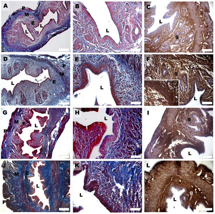
Figure 4.
Mallory Trichrome (A,B,D,E,G,H,J,K) and Col1 (C,F,I,L) staining of mouse uterine horns of control (A–C), DHEA (D–F), DHEA/LC-ALC (G–I) and DHEA/LC-ALC-PLC (J–L) groups. (A,D,G,J) low-magnification LM pictures of the lumen (L), endometrium (E), myometrium (M) and perimetrium (P). (B,E,H,K) high magnification of the luminal epithelium. (C,F,I,L) glandular epithelium (g). Inset in F shows a detail of the tunica muscularis. LM, mag. 10× ((A,D,G,J) Bar: 200 µm), 20× ((B,C,E,F,H,I,K,L) Bar: 100 µm).

