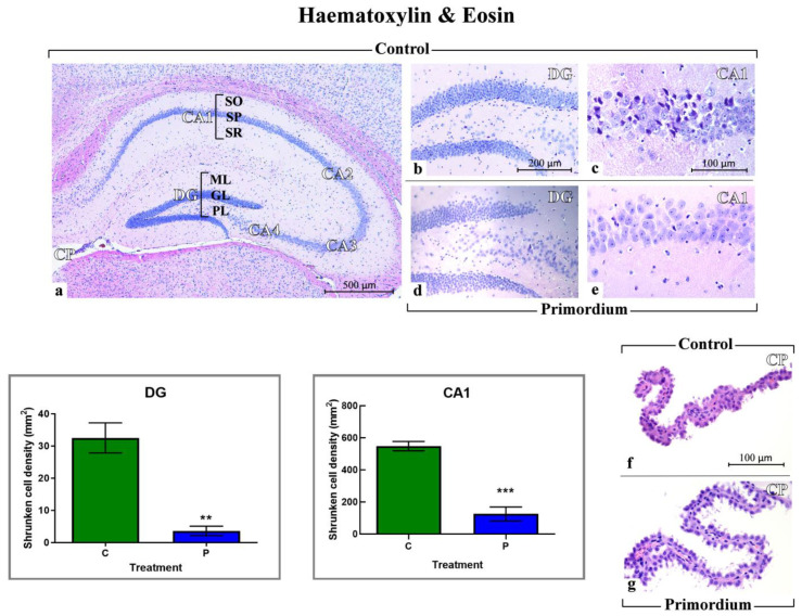
Figure 3.
Histological characterization by H&E staining. Representative brain sections, showing the well-preserved physiological hippocampal cytoarchitecture both in non-supplemented controls (a–d) and P (d,e) aged mice. (a): low magnification micrograph shows the whole hippocampus, formed of cornu Ammonis (CA) and dentate gyrus (DG). CA is further partitioned into: CA1, CA2, CA3 and CA4. The choroid plexus (CP) of the lateral ventricle can be also observed. (b,d): higher magnifications of the DG area revealing well-defined three layers: molecular layer (ML), granule cell layer (GL) and pleomorphic layers (PL). (c,e): higher magnifications of the CA1 region, showing the typical three layered-structure. Outer polymorphic layer, i.e., Stratum oriens (SO); middle pyramidal cell layer, namely Stratum pyramidale (SP); inner molecular layer, i.e., Stratum radiatum (SR). (f,g): choroid plexus (CP) in C and P mice, respectively. (f): evident structural alterations were observable, with ependymal cells displaying cilia reduction. Light microscopy magnification: 40× (a), 200× (b,d), 400× (c,e–g). Scale bars: 500 µM (a); 200 µM (b,d); 100 µM (c,e–g). Lower left panels: Histograms showing the quantitative assessment of shrunken cell density in DG and CA1 region of Ammon’s horn. p values calculated by unpaired Student’s t-test: p < 0.01 (**), and p < 0.001 (***).

