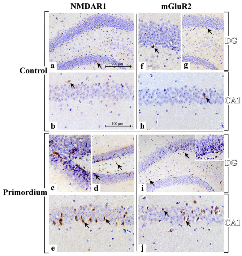
Figure 8.
Immunohistochemical labelling for NMDAR1 and mGluR2 in DG and CA1 region from C animals ((a,b,f–h), respectively) and P ((c–e,i,j), respectively) mice. NMDAR1: sporadic immunolabelled cells (arrows) are observable both in DG (a) and CA1 region (b) of C animals. A marked immunopositivity is clearly evident both in the DG and CA1 area of P mice ((c–e), respectively). In the DG, clusters of heavy immunoreactive cells, arranged in well-ordered chains, are observable in the width of the GL (c), several immunolabelled cells are in the SGZ (d, arrows), and many immunoreactive neurons with large soma are detectable in the PL ((d), arrows). In the CA1 area several NMDAR1-immunopositive neurons are visible localized in the SP, often showing palely immunomarked tiny prolongations, deepening in the underneath SR ((e), arrows). mGluR2: a weak and sporadic immunoreactivity is observable both in DG (f,g) and CA1 region (h) of C animals, where rare immunolabelled cells are detected (arrows). A heavy immunopositivity is clearly evident both in the DG and CA1 area of P mice ((i,j), respectively). Some heavily immunoreactive cells are evident in the DG (i), principally localized in the width of the GL and close to the SGZ (arrows). Various immunomarked neurons are also noticeable in the PL ((i), arrows). Numerous immunopositive neurons are detectable in the SP of CA1, often characterized by immunoreactive prolongations, deepening beneath in the underlying SR (j). Light microscopy magnification: 200× (a,d,g,i); 400× (b,e,h,j); 600× ((c,f) and insert in (i)). Scale bars: 200 µM (a,d,g,i); 100 µM (b,c,e,f,h,j).

