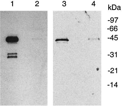
FIG. 6.
Detection of MGT in S. aureus cells. S. aureus cells (0.7 g) grown to log phase were collected and lysed by sonication. The whole-cell lysate was centrifuged at 75,000 × g for 30 min to generate a supernatant fraction (lanes 2 and 4) and particulate fraction (lanes 1 and 3). After separation by SDS-PAGE, Western blotting was performed using either polyclonal antibodies against purified His-ΔMGT (lanes 1 and 2) or preimmune serum (lanes 3 and 4) as described in the text.

