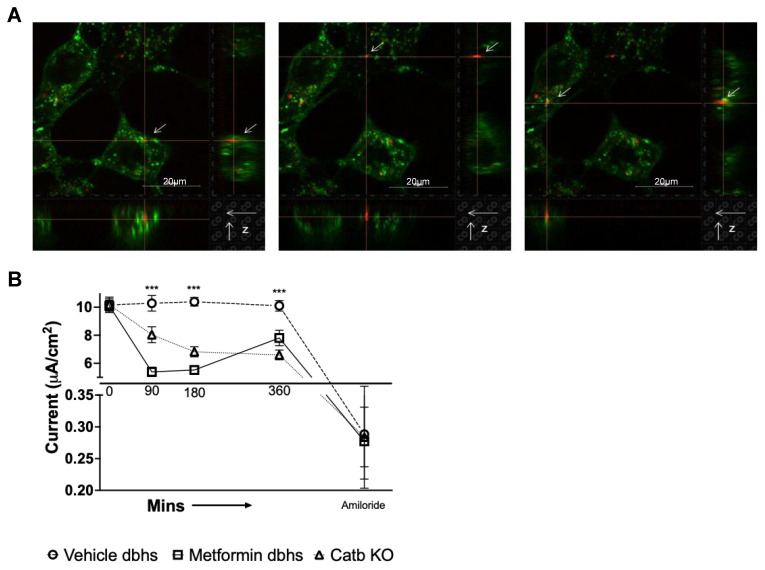
Figure 7.
Uptake of urinary EVs from diabetic db/db mice by mouse mpkCCD cells and changes in amiloride-sensitive transepithelial current after mpkCCD cells were challenged with urinary EVs from vehicle- or metformin-treated db/db mice on an HSD. (A) Immunofluorescence images of mpkCCD cells (green, orthogonal view) treated for 15 min with red fluorescently labeled urinary EVs isolated from diabetic db/db mice. (B) Amiloride-sensitive transepithelial current measurements showing transepithelial current in mpkCCD cells over time after treating the cells with urinary EVs from either diabetic db/db mice maintained on an HSD and treated with vehicle (vehicle) or metformin (metformin), or cathepsin B knockout mice (Catb KO). N = 6 inserts per group, and *** represents a p-value < 0.001. Red fluorescence shows urinary EVs labeled with Claret far red. Green fluorescence shows mpkCCD cells incubated with 1μg/mL of cholera toxin subunit B Alexa Fluor 488 conjugate.

