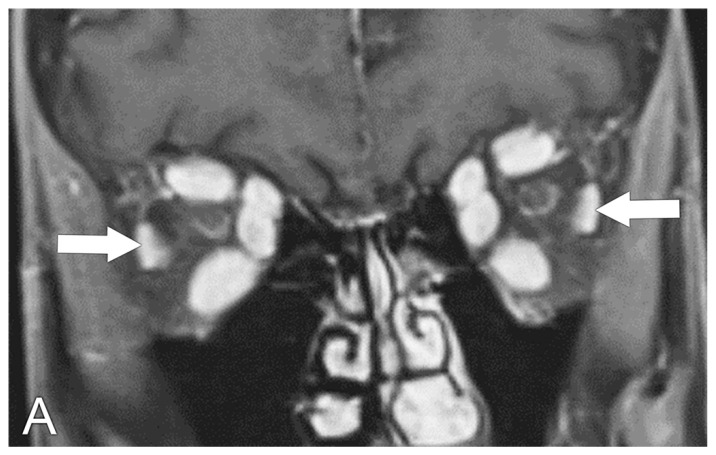
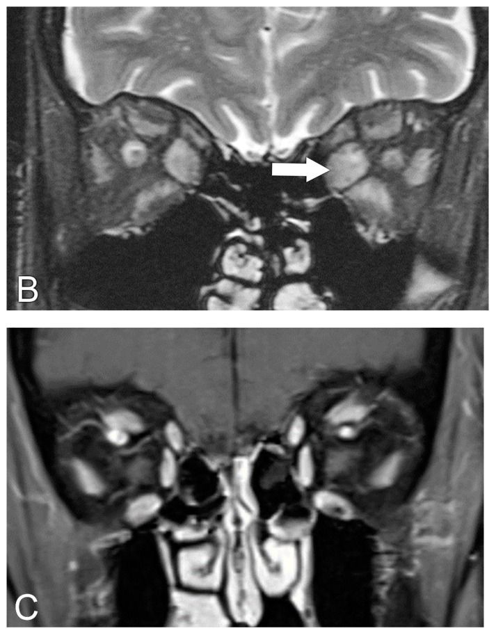
Figure 2.
Graves’ eye disease, MRI. (A) Coronal T1 post-contrast MRI sequence shows enlargement of bilateral extra-ocular muscles with sparring of the lateral rectus muscles (arrows). (B) Coronal T2 MRI sequence shows hyperintense signal signifying edema in the enlarged extraocular muscles (arrow). (C) Coronal T1 post-contrast MRI sequence shows the normal appearance of the extra-ocular muscles.


