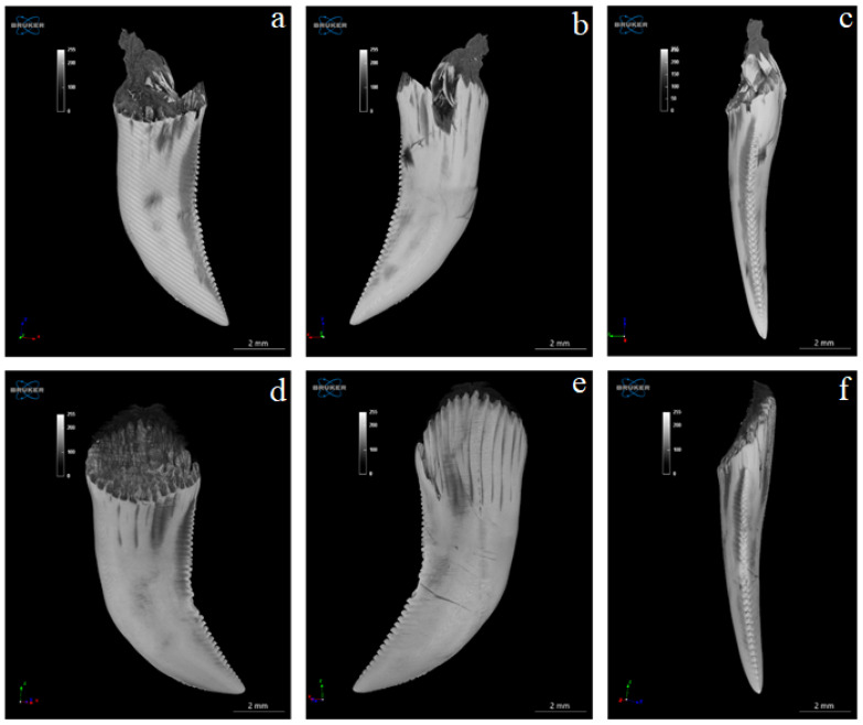
Figure 6.
Microtomography analysis of Komodo dragon (Varanus komodoensis) 2 teeth (a–f) in a different orientation. (a,b)—both surfaces (vestibular and lingual) of the same tooth and (c)—its distal edge. (d,e)—both surface (vestibular and lingual) of the second tooth and (f)—its distal edge. Shallow grooves visible in the apical part of the tooth on its mesial margin and deeper, numerous grooves present on the distal margin of the tooth. Note the carina of the distal margin of the tooth (a–f).

