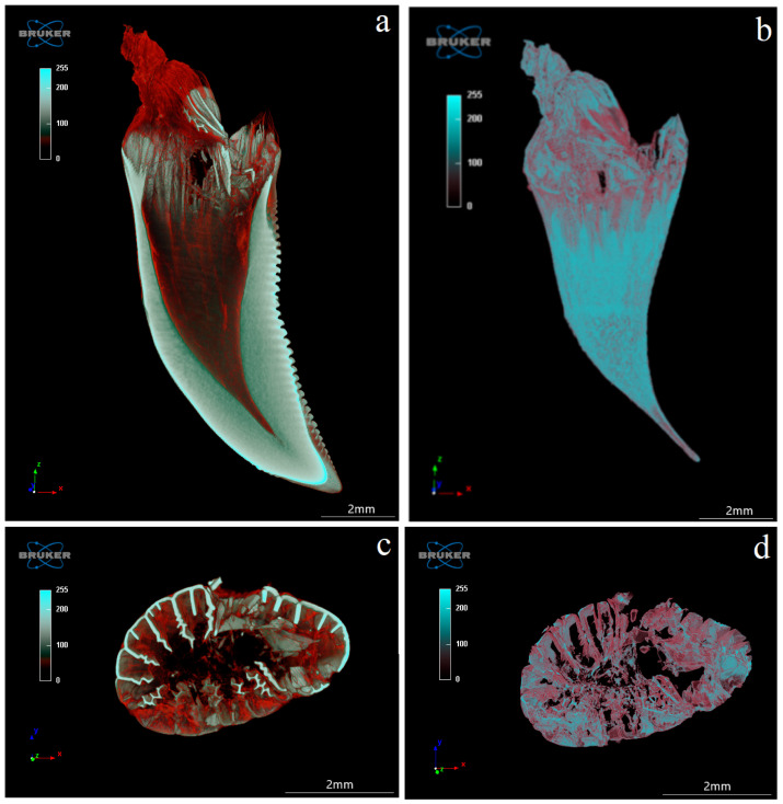
Figure 12.
Coronal (X-Z) (a,b) and transaxial (X-Y) (c,d) sections of the mandibular tooth of the Komodo dragon (Varanus komodoensis). (a)—connective tissue and blood vessels stained in red, adjacent to the dentine (white); (b)—tooth cavity with connective tissue and blood vessels stained in red; (c)—note plicidentine producing numerous secondary lamellae, and blood vessels stained in red; (d)—connective tissue with blood vessels. Scale bar: 2 mm.

