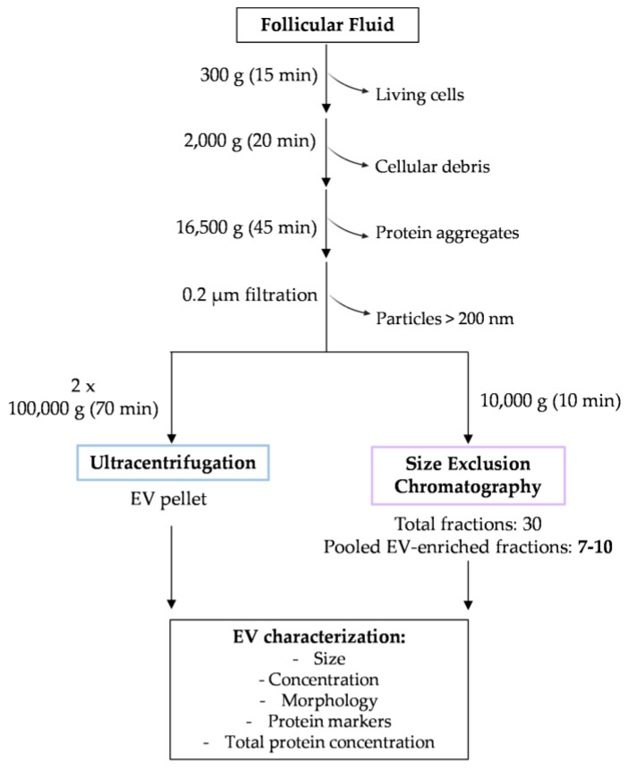
Figure 1.
Schematic representation of the experimental design. FF samples were centrifuged at 300× g and 2000× g to exclude living cells and cellular debris, respectively. The supernatant was centrifuged at 16,500× g to pellet protein aggregates. A 0.2 µm filtration step was conducted to remove particles larger than 200 nm. From this volume, 1 mL was stored at −80 °C for EV isolation using SEC, and the remaining volume was ultracentrifuged twice to isolate the final UC EV pellet. In total, 30 fractions of 500 µL were collected, and EV-enriched fractions (F7–10) were used for further characterization. The EVs isolated using UC and SEC F7–10 fractions were characterized regarding the particle size, concentration, morphology, presence of specific markers, and the total protein content using DLS, NTA, TEM, Western blot, and microBCA, respectively.

