Figure 2.
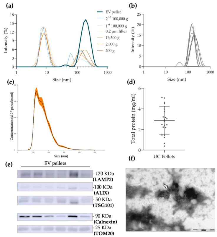
Characterization of extracellular vesicle (EV) samples isolated using ultracentrifugation (UC): (a) dynamic light scattering (DLS) analysis of particle size distribution in each supernatant of differential centrifugation protocol and EV final pellet; (b) DLS analysis of particle size distribution in follicular fluid (FF)-derived UC pellets (n = 9); (c) nanoparticle tracking analysis (NTA) of particle concentration and size distribution in FF-derived UC pellets (n = 3; orange bars indicate ± SEM); (d) total protein quantification of each EV pellet using microBCA kit (n = 22); (e) Western blot membrane showing the expression of specific EV positive markers, such as LAMP2, ALIX, and TSG101, and also the presence of cellular contaminants in the final UC pellet, such as Calnexin and TOM20 (n = 6; see Supplementary materials for controls); (f) representative image of EVs isolated using UC obtained with transmission electron microscopy observations (EV cup-like structure indicated by the arrow).

