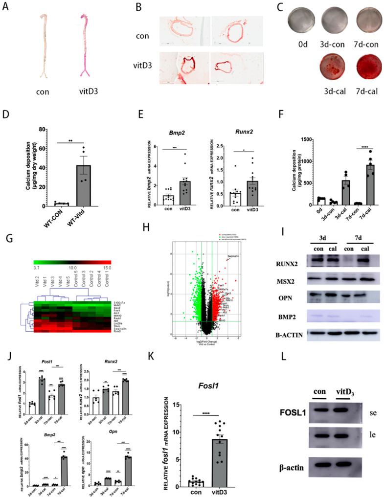
Figure 1.
FOSL1 is up-regulated in vascular calcification both in vivo and in vitro. (A) Representative ARS staining of whole aortic arteries isolated from C57 mice (original magnification scale bars =), n = 3 per group. (B) Mouse arteries were isolated from vitD3 animal models, mineral deposition in the aortic section was detected by ARS staining. (C) Calcium content of arteries was measured by a calcium assay kit, n = 5 independent experiments. (D) Calcium deposition in aortas was detected by ARS staining.; n = 4 per group. (E) mRNA expression levels of osteogenesis genes in animal aortas were analyzed by RT-qPCR, n = 12 per group. (F) Mouse vascular smooth muscle cells (VSMCs) were incubated in the presence of calcifying medium for seven days. Mineral deposition in VSMCs was detected by ARS staining, n = 5 per group. (G) Heatmap of differentially expressed genes, n = 5. (H) Volcano plot showing FOSL1 as one of the most upregulated genes in microarray analysis: log 2-fold change versus log 2-fold regulation, n = 50 per group. (I) Western blot analysis of RUNX2, MSX2, OPN, and BMP2 protein expression, n = 3 per group. (J) Quantitative real-time polymerase chain reaction analysis of FOSL1, BMP2, RUNX2, and OPN mRNA expression in mouse VSMCs, n = 5 per group. (K) FOSL1 mRNA expression in animal arteries, n = 12 per group. (L) FOSL1 protein expression in mouse arteries, n = 3 per group. Statistical analysis was performed using one-way ANOVA (Tukey honestly significant difference post hoc test) * p < 0.05, ** p < 0.01, *** p < 0.001, **** p < 0.0001.

