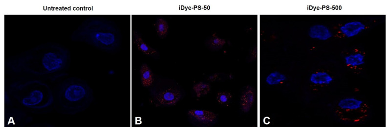
Figure 3.
The confocal images depict the internalization of iDyePS-50 and iDyePS-500 in HNEpCs after exposures (100 µg/mL) lasting for 24 h. (A) Untreated control. (B) Localization of iDyePS-50 (in red) in cytoplasmic regions and surrounding nuclei (blue). (C) Presence of iDyePS-500 (in red) in the cytoplasm and proximity to nuclei.

