Figure 1.
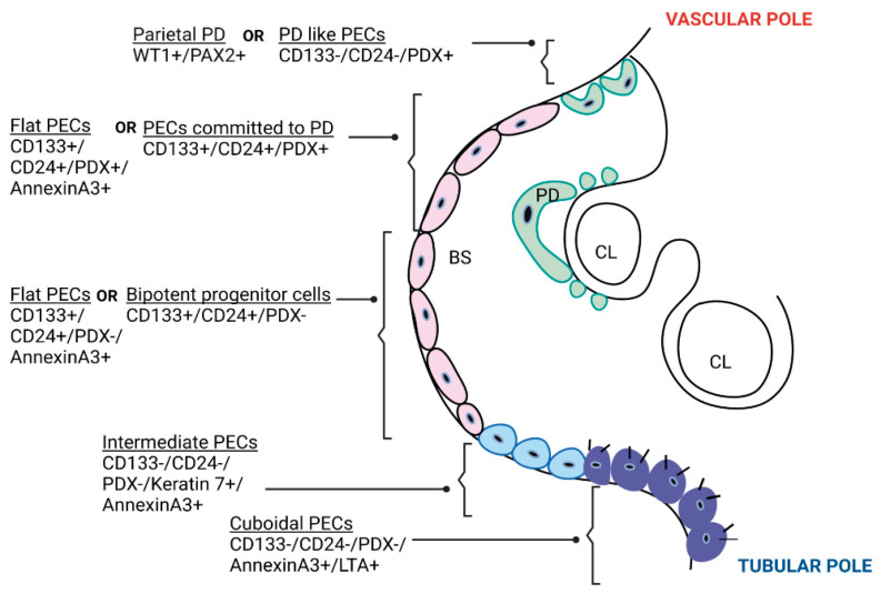
Types of parietal epithelial cells. A cartoon showing transverse section of a glomerulus showing types of PECs based on location, molecular phenotype [3,20] and morphology [21]. PECs based on stem cell marker (CD133/CD24) and PDX expression were located hierarchically along the Bowman’s capsule; differentiated cells resembling PD (CD133-CD24-PDX+) were located at the vascular pole, progenitor cells committed to PD (CD133+CD24+PDX+, also called “transitional cells”) were located between the vascular pole and tubular pole, and bipotent progenitor cells (CD133+CD24+PDX-, with a high regenerative capacity towards both PD and proximal tubular cells) were located at the tubular pole [3]. PECs based on morphology were: flat squamoid cells, intermediate PECs, and cuboidal PECs [21]. PD—podocyte; PECs—parietal epithelial cells; CL—capillary lumen; BS—Bowman’s space; WT1—Wilms’ tumor protein 1; PAX2—paired box gene 2; CD—cluster of differentiation; PDX—podocalyxin; LTA–lotus tetragonolobus agglutinin; FSGS–focal segmental glomerulosclerosis; CrescGN–crescentic glomerulonephritis.

