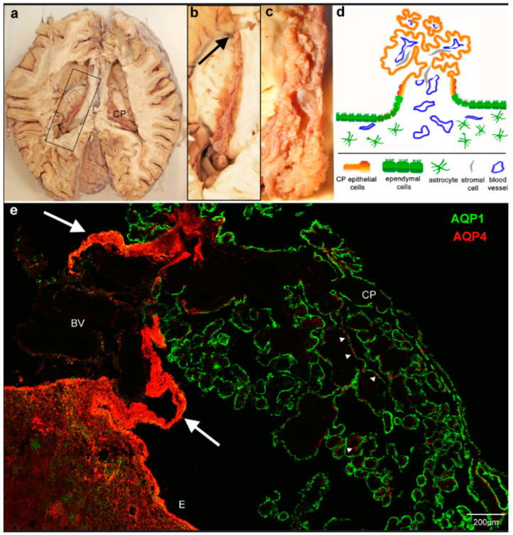
Figure 1.
Overview of the location and attachment of the choroid plexus and aquaporin expression. (a) Horizontal section through a human brain with open lateral ventricles. (b) View of the boxed area indicated in a. showing the choroid plexus (CP) from the anterior end close to the interventricular foramen (arrow) to the most posterior part, where it thickens to the glomus and extends further into the lower horn of the lateral ventricle. (c) Detailed view of the posterior region shows the firm attachment to the thalamic surface. (d) The schematic diagram of a cross-section perpendicular to the plane shown in c indicates the direct transition of the ependyma to the choroid plexus epithelium. (e) Cryostat section through the choroid plexus and ependymal attachment of the lateral ventricle stained for aquaporin 1 and 4 (AQP1, AQP4). CP epithelium is positive for AQP1, astrocytic endfeet in the brain parenchyma and ependyma (E), and some cells (arrowheads) in the CP are positive for AQP4. Note that the transitional ependyma connecting to the CP epithelium and underlying tissue is strongly immunofluorescent for AQP4 (arrows). BV, blood vessels.

