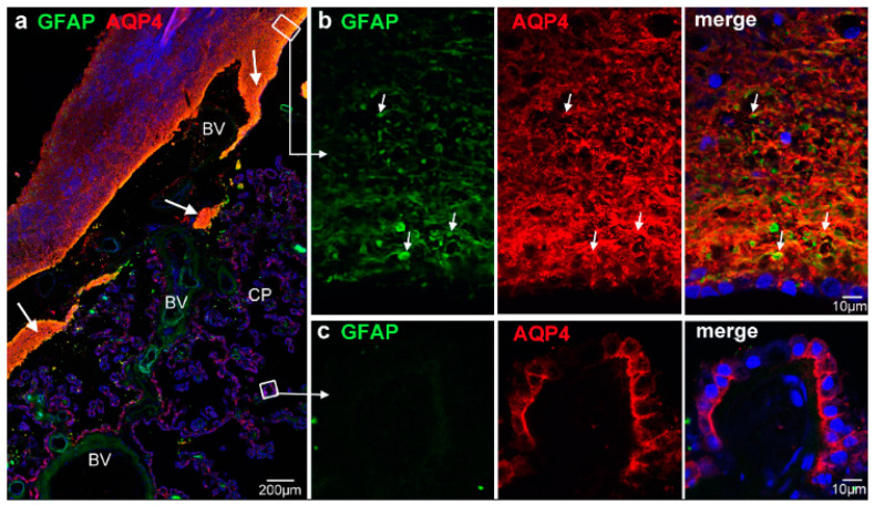
Figure 2.
Subependymal tissue in the transitional zone is formed by astrocytes. (a) At low magnification, GFAP immunoreactivity largely overlaps with AQP4 stain under the transitional ependyma (large arrows). (b). Detailed view from the ependymal/subependymal region indicated by the white box shows a dense meshwork of GFAP-positive processes and strong AQP4 immunoreactivity. The GFAP processes are often surrounded or associated with AQP4 staining (small arrows). The surface ependymal cells are only weakly positive for GFAP. (c) GFAP stain is completely lacking in AQP4-positive CP epithelial cells. Nuclei are stained with DRAQ5. BV, blood vessels.

