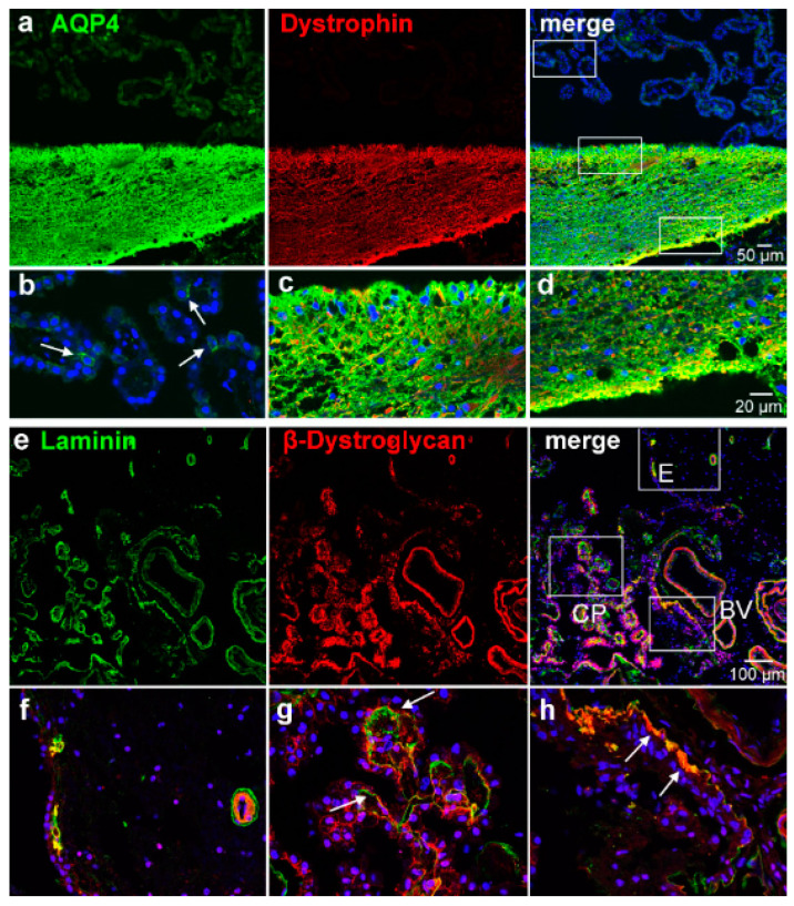
Figure 4.
Dystrophin and β-dystroglycan localization in the CP and ependymal transition zone. (a) Co-staining with antibodies against AQP4 and dystrophin shows an overlap in the glial subependymal plate but not in the CP. The white rectangles indicate the details shown (b–d). (b) AQP4-positive cells in the CP (arrows) are negative for dystrophin, whereas (c,d) subependymal astrocytic processes were intensely positive for both dystrophin and AQP4. An even stronger immunoreactivity is found on the perivascular side where the processes face a basal lamina than on the ependymal side of the glial plate (d, c.f., Figure 3b,f). (e) β-dystroglycan and laminin co-stainings show a close association of both stains. Detailed views (f–h) of the indicated areas reveal that the ependyma as well as astrocytic processes in the glial plate were immunonegative for β-dystroglycan (f), except where they contact a basal lamina as indicated by laminin staining. Both stains are also found in and around blood vessel walls (h). In the choroid plexus (CP), epithelial cells express β-dystroglycan basolaterally close to the basal lamina (g).

