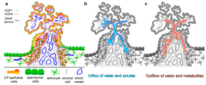
Figure 5.
Summary and suggested implications of aquaporin expressions in the ependyma–choroid plexus transition zone. (a) AQP4 expression is particularly high in astrocytic processes that form a glial plate or cuff around the blood-supplying vessels of the CP. AQP4 staining is also found basolaterally in some CP epithelial cells (red lines). The astrocytic processes delimited by a basal lamina reach the CP stroma, which includes fenestrated capillaries. AQP1 is expressed mostly apically in CP epithelial cells, which also form tight junctions as the basis for the blood–CSF barrier. Note that the basal lamina of blood vessels is not included in the schematic. (b) Water and solutes can diffuse into the CP stroma through fenestrated capillaries and can enter the brain parenchyma via the glial plate. The basal laminae might have a filtering effect but do not constitute a barrier. Water flow is likely restricted by the dense meshwork of astroglial processes in the glial plate. (c) Depending on the osmotic gradient, the high density of AQP4 water channels in the glial plate could also serve as a drainage and clearing pathway for metabolites, which are then taken up by postcapillary venules in the stroma. This would contribute significantly to processes suggested by the glymphatic pathway hypothesis.

