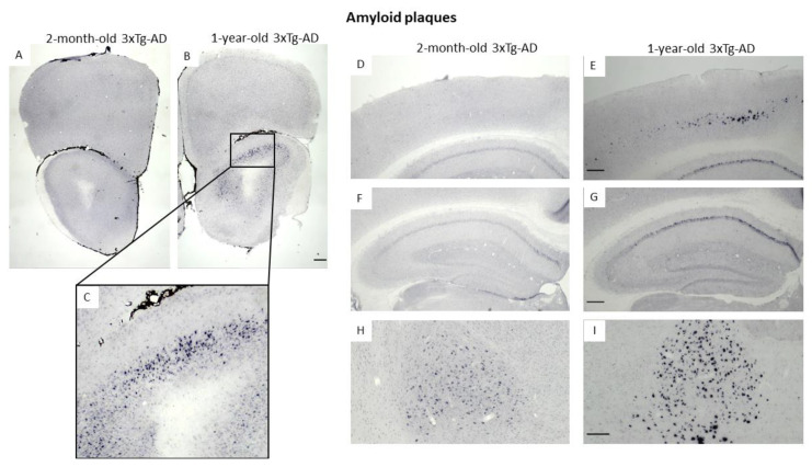
Figure 6.
Amyloid-β1–42 plaques stained with Ni-DAB immunohistochemistry in 2-month-old and 1-year-old 3xTg-AD mice. The images show different brain areas: the olfactory bulb (A–C), motor- and somatosensory-cortex, (D,E), hippocampus (F,G), basolateral and basomedial amygdala (H,I). Scale bar in (B,E,G) is 200 μm, and in (I) 100 μm.

