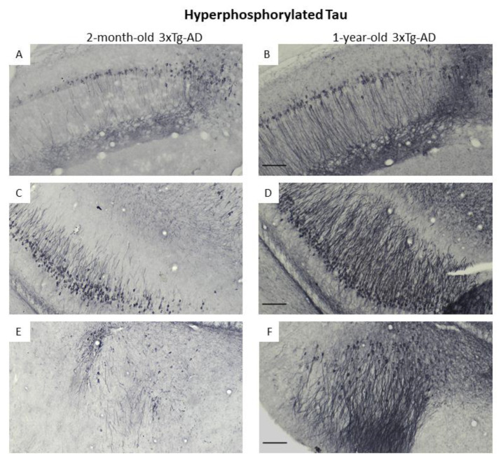
Figure 7.
Phospho-Tau tangles stained with Ni-DAB immunohistochemistry in 2-month-old and 1-year-old 3xTg-AD mice. The images show different brain areas, like: pyramidal cell layer in the CA1 region of the hippocampus (A,B), pyramidal cell layer in the CA3 region of the hippocampus (C,D) and basolateral amygdaloid nucleus, amygdalopiriform area and entorhinal cortex (E,F). Scale bar is 100 μm.

