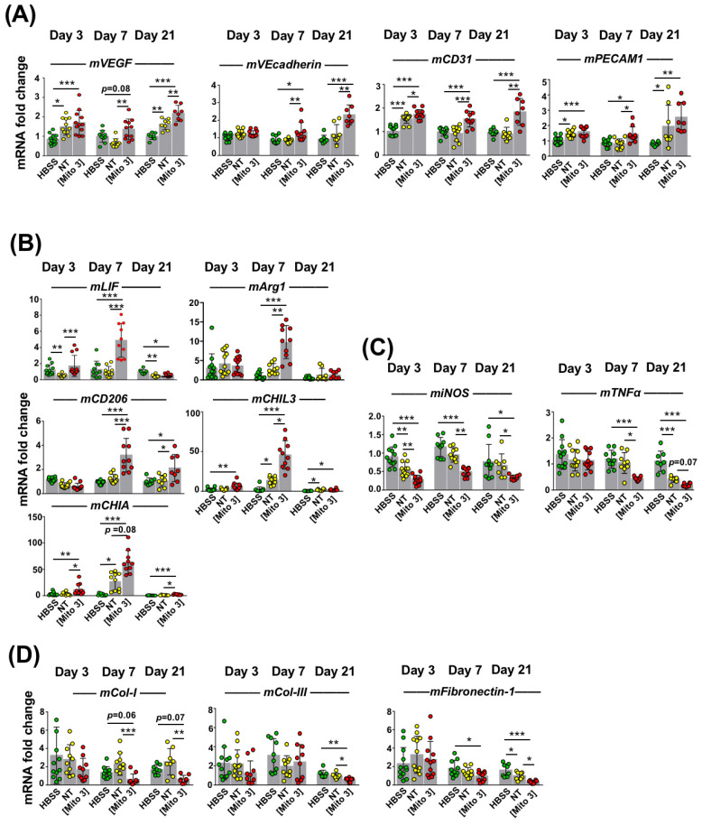
Figure 6.
Cardiac mitochondria transfer improves the pro-angiogenic, anti-inflammatory and anti-fibrotic effects of grafted MSCs. (A–D) MSCs were exposed to cardiac mitochondria at the Mito 3 concentration prior to engraftment in mouse hearts. Relative mouse mRNA expression of (A) VEGF, VEcadherin and CD31; (B) the anti-inflammatory cytokine LIF or the anti-inflammatory M2 macrophage markers (Arg1, CD206, CHIL3 and CHIA); (C) the pro-inflammatory cell marker iNOS and the pro-inflammatory cytokine TNFα; and (D) the extracellular matrix components collagen-1 (Col-1), collagen-3 (Col-3) and fibronectin-1 in infarcted mouse hearts grafted with either human non-treated or mitochondria-preconditioned MSCs in reference to mouse infarcts injected with a saline solution (HBSS) at day 3 (n = 12), day 7 (n = 10), and day 21 (n = 8) post-surgery and graft. One-way ANOVA with Tukey’s multiple comparisons test for mVEGF, mVEcadherin, mCD31, miNOS, mTNFα and mFibronectin-1. One-way ANOVA with Dunn’s multiple comparisons tests were performed for the other genes. * p < 0.05, ** p < 0.01, *** p < 0.001. Each dot represents a mouse. Bar graphs represent mean values ± SD.

