Figure 7.
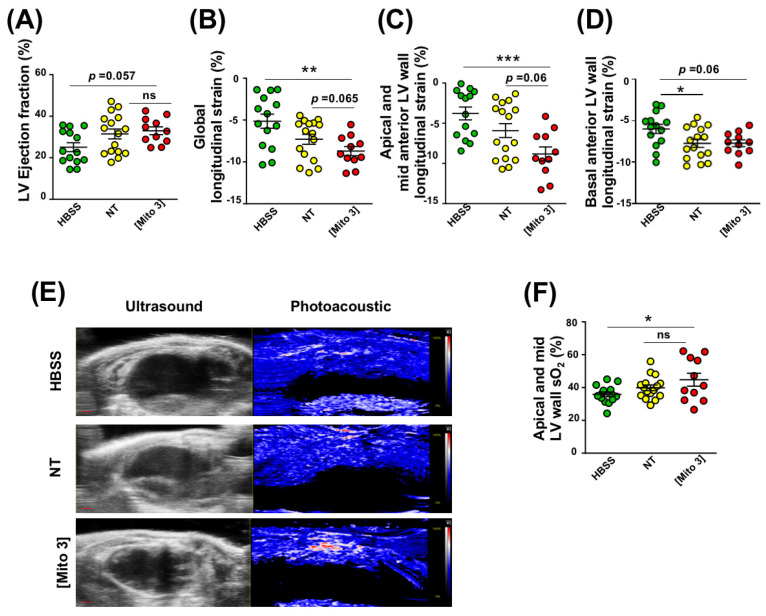
Cardiac mitochondria transfer enhances cardiac function and myocardial perfusion 21 days after myocardial infarction. (A–D) MSCs were conditioned with cardiac mitochondria (Mito3 concentration) 24 h prior to engraftment in infarcted mouse hearts. Different cardiac functional parameters were evaluated in infarcted mice treated with a saline solution (HBSS), non-treated MSCs (NT), or cardiac mitochondria-preconditioned MSCs (Mito 3) at day 21 post-surgery and graft. (A) LV ejection fraction; (B) global longitudinal strain; (C) apical and mid anterior LV wall longitudinal strain; (D) basal anterior LV wall longitudinal strain. (E) Representative B-mode ultrasound parasternal long-axis view to define the left ventricular anterior wall and photoacoustic mode with color scaling to show areas of high oxygen saturation in red and low oxygen saturation in blue in hearts from the different mouse treatment groups. Scale bar: 1 mm. (E) Variation of myocardial anterior wall sO2 in the different mouse treatment groups. One-way ANOVA with Tukey’s multiple comparisons test in (A–D,F). * p < 0.05, **p < 0.01, *** p < 0.001. Each dot represents a mouse. Infarcted mice treated with a saline solution (HBSS) (n = 11), non-treated MSCs (NT) (n = 11), or cardiac mitochondria-preconditioned MSCs (n = 10). Bar graphs represent mean values ± SEM.

