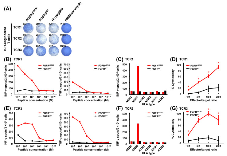
Figure 3.
Functional assays of FGFR3Y373C-specific TCR-engineered T cells. (A) IFN-γ ELISPOT assay on FGFR3Y373C-specific TCR-engineered T cells generated by retroviral transduction of healthy donor’s PBMCs with TCRs. (B,E) Secretion of cytokines, IFN-γ (left) and TNF-α (right), of FGFR3Y373C TCR-engineered T cells of TCR1 (B) and TCR3 (E) stimulated by C1R-A0206 cells loaded with graded amounts (10−5 to 10−10 M) of FGFR3Y373C or wild-type FGFR3WT peptides. Data are represented as means in duplication experiments. (C,F) HLA-restricted responses of FGFR3Y373C-specific TCR-engineered T cells expressing TCR1 (C) and TCR3 (F). IFN-γ ELISPOT assay of FGFR3Y373C-specific TCR-engineered T cells stimulated by co-culturing with C1R-A0201, C1R-A0206, C1R-A1101, C1R-A2402, C1R-A3101, and C1R-A3303 cells loaded with FGFR3Y373C or FGFR3WT peptide. (D,G) Cytotoxic activity of FGFR3Y373C-specific TCR-engineered T cells of TCR1 (D) and TCR3 (G) against C1R-A0206 cells pulsed with FGFR3Y373C or FGFR3WT. The cytotoxic activity was measured in different ratios (1:1, 5:1, 10:1, and 20:1) of FGFR3Y373C-specific TCR-engineered T cells (effector cells) and peptide-loaded C1R-A0206 cells (target cells). Data are represented as means with standard deviations in quadruplicate experiments. Asterisks indicate statistically significant differences (p < 0.05) between the two groups.

