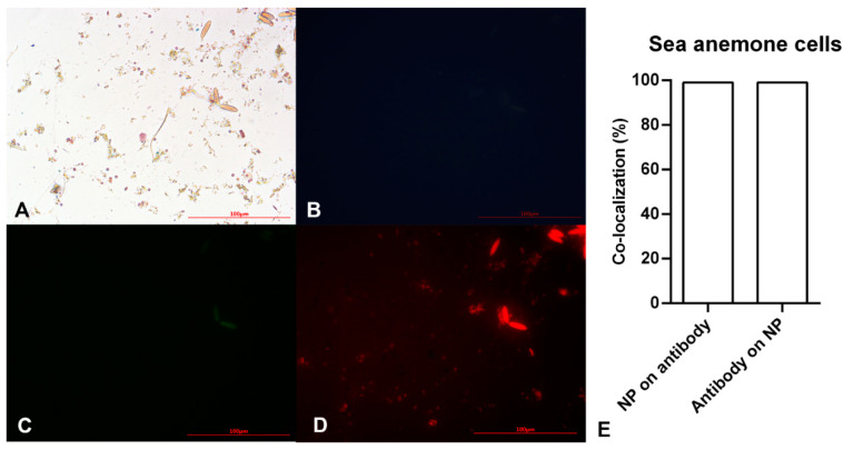
Figure 4.
Microscopy images of sea anemone stem cells after exposing to Alexa Flour anti-Oct4 antibody-conjugated iron nanoparticles using (A) brightfield, (B) blue filter, (C) green filter, and (D) red filter. (E) Co-localization of conjugated iron nanoparticle staining and anti-Oct4 antibody in sea anemone stem cells. Columns show Manders’ coefficients expressed as percent values.

