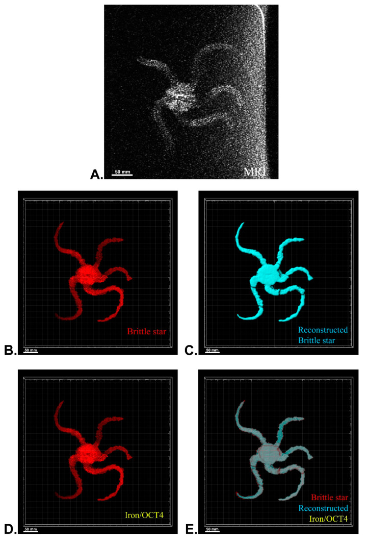
Figure 6.
Three-dimensional (3D) reconstruction of brittle star tissue with no treatment using magnetic resonance imaging (MRI) serial images: (A) original grayscale image; (B) whole tissue imaging and signals of the brittle star tissue without reconstruction; (C) brittle star whole tissue 3D reconstruction in which the blue surface represents reconstructed brittle star tissue; (D) 3D reconstruction of the iron/iron plus anti-Oct4 signals, which is negative in this image; (E) 3D reconstruction of the whole tissue of brittle star along with all signals. All scale bars in four images are 50 mm. All images were processed and generated by Imaris software (V 7.4.2, ImarisX64; Bitplane AG, Schlieren, Switzerland).

