Figure 5.
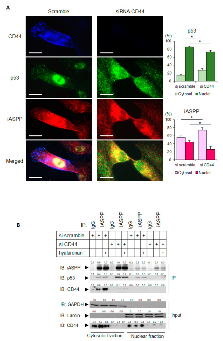
Depletion of CD44 promotes the translocation of p53 and iASPP to the cytoplasm. (A) hTERT-BJ cells treated with CD44 siRNA, or not, were stained for CD44 (blue), p53 (green), or iASPP (red). Images were taken using a Zeiss Axioplan 2 immunofluorescence microscope with a 63× objective. By ImageJ software, the localizations of p53 and iASPP in cytosol and nuclei were measured and are shown in the panels to the right. Scale bars, 10 µm. * p < 0.05, Student´s t-test. (B) Subcellular fractionation was performed on untreated or hyaluronan-stimulated hTERT-BJ cells transfected with siRNA for CD44 or scramble siRNA as control. Cell lysates were then immunoprecipitated with an iASPP antibody, followed by immunoblotting with iASPP, p53, and CD44 antibodies. The purity of the nuclear and cytoplasmic fractions was determined by immunoblotting for laminin and GAPDH, respectively. A representative experiment out of three independent experiments performed is shown. The data are plotted in bar graphs representing the mean ± SD of at least three independent experiments (* p < 0.05, Student’s t-test). The uncropped blots are shown in File S1.

