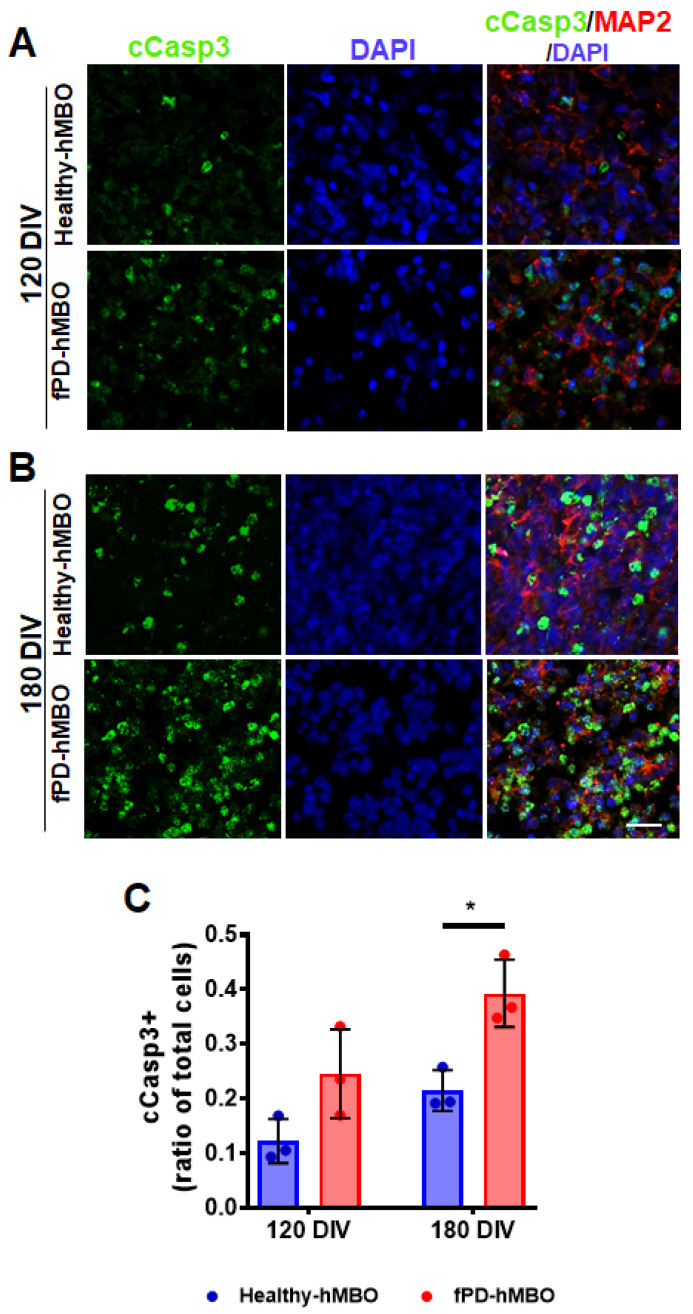
Figure 6.
Elevated apoptosis in fPD-hMBOs. Representative images from healthy hMBOs and fPD-hMBOs, immunostained for c-Casp3 (green) and MAP2 (red), at 120 DIV (A) and 180 DIV (B). Nuclei were stained with DAPI. The scale bar is 25 µm. (C). Quantification of c-Casp3 immunostaining at 120 and 180 DIV. Values represent mean +/− standard error of mean (SEM) from n = 3 hMBOs per group. Three sections were analyzed from each hMBO. The results were analyzed using two-way ANOVA [SNCA copy number: F (1, 8) = 20.13, p = 0.0020; Time: F (1, 8) = 12.65, p = 0.0074); SNCA copy number × time: F (1, 8) = 0.6670, p = 0.4377)] followed by the Tukey post hoc multiple comparisons test. * p < 0.05.

