Figure 2.
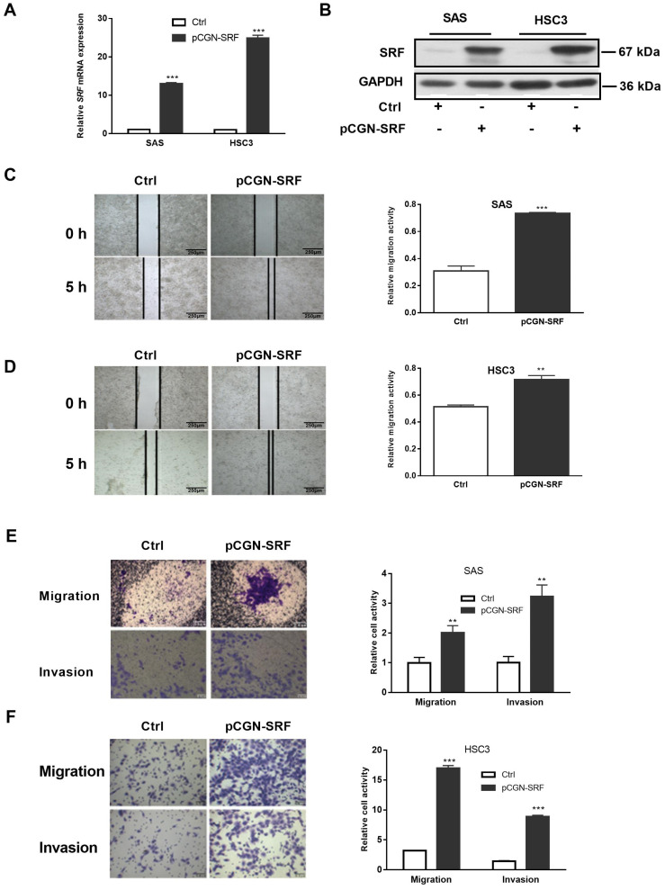
Overexpression of SRF facilitates OSCC cell migration and invasion. Serum-starved OSCC cells (SAS and HSC3) were transfected with pCGN-SRF or control vectors. The expression of SRF mRNA and protein were detected by performing qRT-PCR (A) and western blotting (B), respectively, at 36 h after transfection. The loading control was GAPDH. Original western blots are shown in Figure S5. Wound healing scratch assays of the migration capacity of SAS (C) and HSC3 (D) cells transfected with pCGN-SRF or control vectors. Surfaces of confluent cells were scratched 12 h after transfection. Left, representative images at 0 and 5 h after scratching. Right, quantitation of cell migration capability. Scale bar: 250μm. Migration and invasion capacity of SAS (E) and HSC3 (F) cells transfected with pCGN-SRF or control vectors using Transwells. Left, representative images; scale bar: 0.5 μm. Right, quantitation of cell migration and invasion capability. Data are representative of three independent experiments. ** p < 0.01, *** p < 0.001 vs. control. Ctrl, control; OSCC, oral squamous cell carcinoma; qRT-PCR, quantitative reverse transcription-polymerase chain reaction; SRF, serum response factor.

