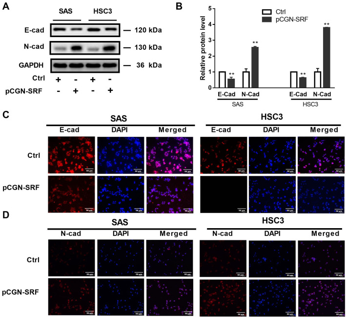
Figure 3.
Serum response factor promotes OSCC cell epithelial-to-mesenchymal transition. Serum-starved OSCC cells (SAS and HSC3) were transfected with pCGN-SRF or Ctrl vectors. At 72 h post-transfection, the protein levels of E-cadherin and N-cadherin were detected via western blotting. GAPDH served as a protein loading control (A). Original western blots are shown in Figure S6. The relative band intensity was determined through densitometric analysis (B). Data are presented as the mean ± SD of three independent experiments. ** p < 0.01 vs. control vector. (C) Immunofluorescence staining of E-cadherin in SAS and HSC3 cells transfected with pCGN-SRF or Ctrl vectors. (D) Immunofluorescence staining of N-cadherin in SAS and HSC3 cells transfected with pCGN-SRF or control (Ctrl) vector. Scale bar: 50μm. Data are representative of three independent experiments. Ctrl, control; E-cad, E-cadherin; N-cad, N-cadherin; OSCC, oral squamous cell carcinoma.

