Figure 4.
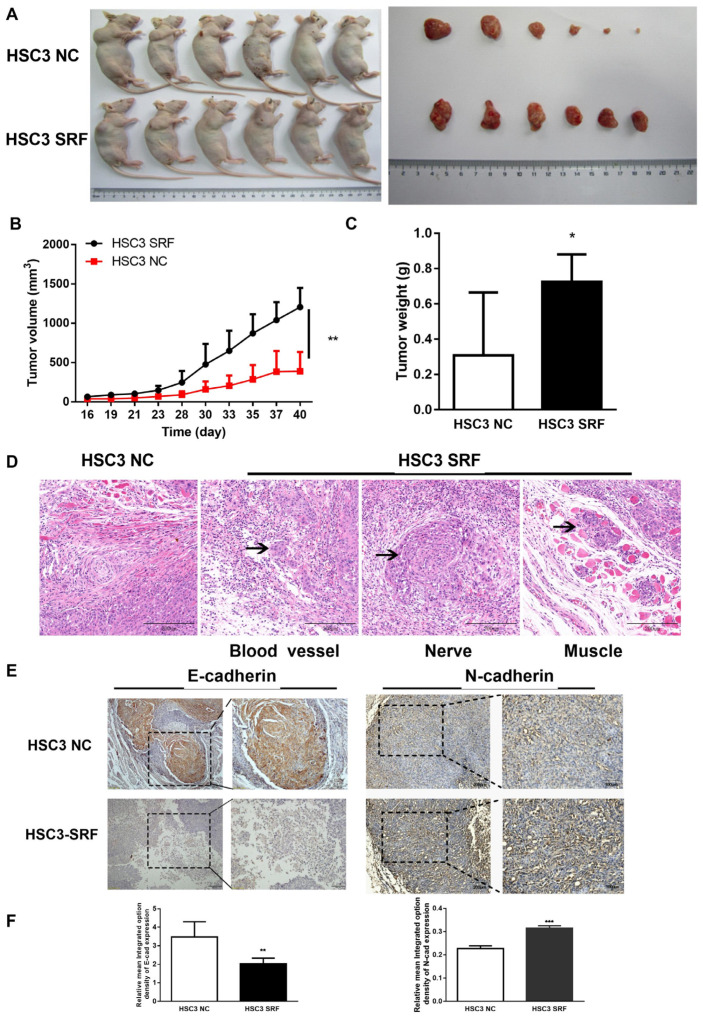
SRF overexpression promotes OSCC tumorigenesis in vivo. Xenografted nude mice (n = 6/group) were injected with HSC3 cells stably overexpressing SRF. The mice were euthanized, and tumors were excised from nude mice 40 days after HSC-3 cell injection (A). Tumor volumes were measured using calipers (B), and tumors were weighed at the end of the experiment (C). Statistical data were derived from three independent measurements and are shown as means ± SDs. Infiltration of HSC-3-SRF tumor cells (D). Representative immunohistochemical images of E-cadherin and N-cadherin expression in samples from HSC-3-NC and HSC-3-SRF cells (E). The scale bar is shown in lower-right corner. Mean optical density of E-cadherin and N-cadherin in tumor samples from HSC-3-NC and HSC-3-SRF cells (F). * p < 0.05, ** p < 0.01, *** p < 0.001, tumor samples from HSC-3-NC cells compared with HSC-3-SRF cells. NC, negative control; OSCC, oral squamous cell carcinoma; SRF, serum response factor.

