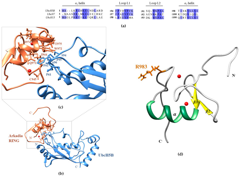
Figure 9.
(a) Sequence alignment of helix a1, loop L1, loop L2, and helix a2 of UbcH5B, UbcH7, and UbcH13 E2 enzymes, showing the amino acid conservation among the interacting with Arkadia regions. (b) Model of the UbcH5B–Ark RING complex (UbcH5B in light blue and Ark RING in coral). The model was prepared by superimposing Ark RING (PDBid: 2KIZ) to the X-ray structure of the UbcH5B-c-CBL complex (PDBid: 4A49) and extracting the c-CBL structure. Zn(II) ions are depicted as red spheres. (c) Close-up of the theoretical UbcH5B–Ark RING interface highlighting the central role of UbcH5B’s Phe62 and Ser94-Pro95-Ala96 motif. Key contact residues from Arkadia and UbcH5B are shown as coral and light blue sticks, respectively. (d) NMR structure of Ark RING (PDBid: 2KIZ) with Arg983 shown in orange sticks.

