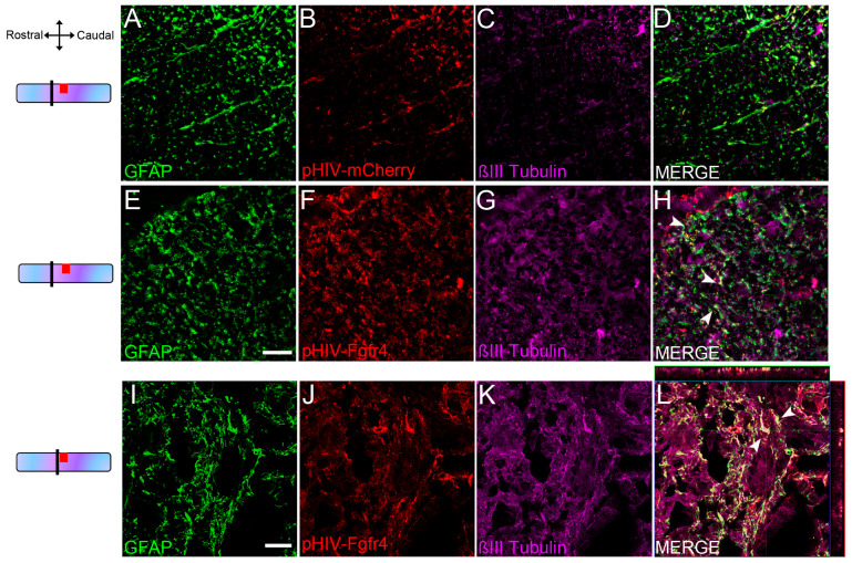
Figure 2.
Transduced astrocytes express βIII-tubulin 6 weeks after SCI. Schemes on the left indicate the location of images. Representative THUNDER images of pHIV-mCherry-injected female mice (dorsal white matter) 2 mm rostral to the lesion at 6 weeks after SCI. Displayed images represent maximum intensity projection of a z stack of 10 planes with a 3 μm thickness (A–D). Representative THUNDER images of pHIV-Fgfr4-injected female mice (dorsal white matter) 2 mm rostral to the lesion at 6 weeks after SCI (E–H). Note the increase in βIII-tubulin expression in pHIV-Fgfr4-injected female mice compared to pHIV-mCherry-injected group (A–D). Representative confocal images of pHIV-Fgfr4-injected female mice in close vicinity of the lesion site, 6 weeks after SCI (I–L). Micrographs confirm βIII-tubulin protein expression in a sub-population of transduced astrocytes (arrowheads, (H)). Scale bars: 20 µm.

