Figure 4.
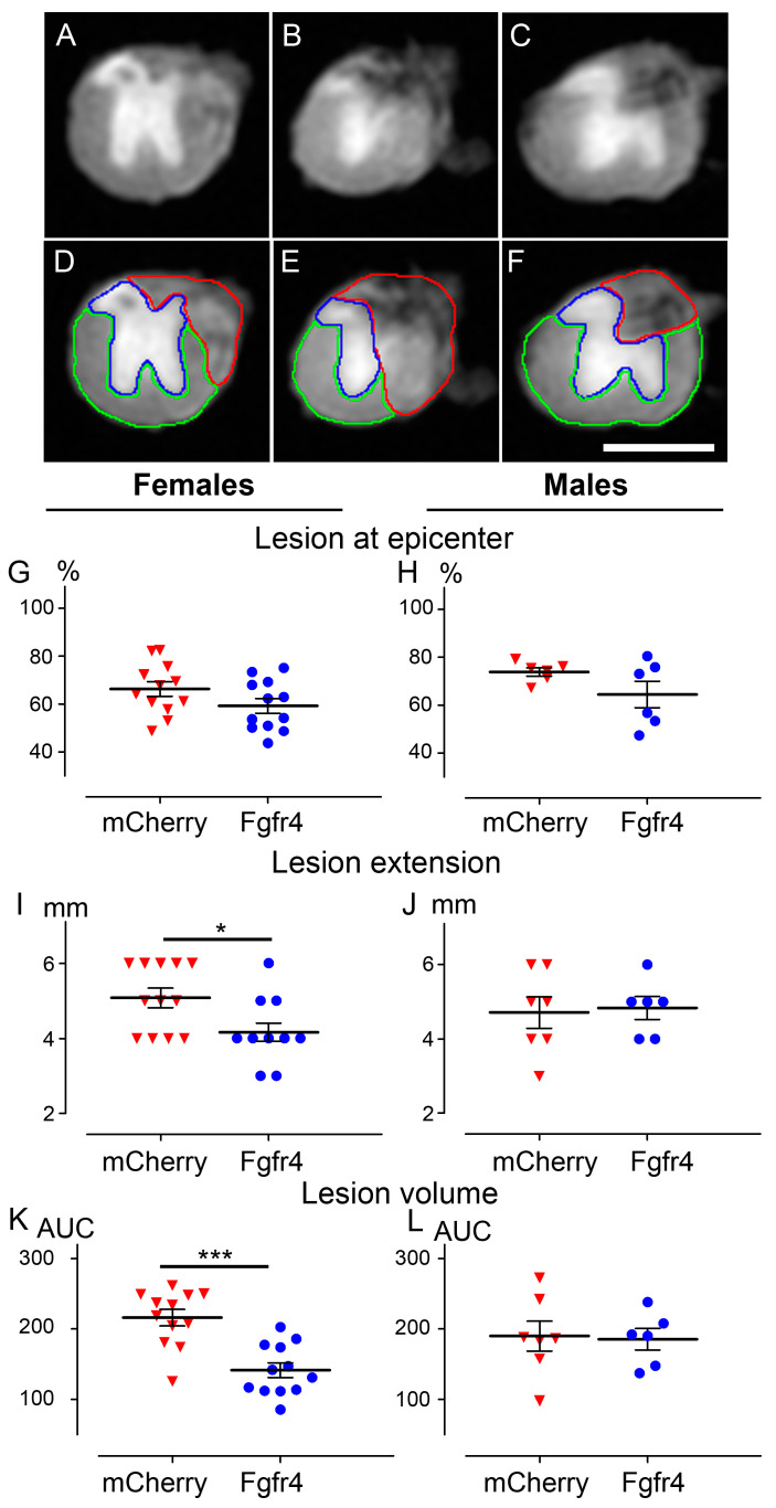
pHIV-Fgfr4 vector injection immediately after SCI preserves spinal cord tissues. Representative ex vivo axial DW-MRI (A–F) of the spinal cord from a C57BL6/6J female mouse that underwent injury and pHIV-Fgfr4 vector injections. Photograph within the lesion epicenter (B,E), 1 mm rostral (A,D), and 1 mm caudal (C,F) to the lesion site. Manual segmentations (D–F) of the spared grey matter (blue), the spared white matter (green), and the injured tissue (red). In females (G,I,K) and males (H,J,L), lesion area at the epicenter (G,H), lesion extension (I,J), and lesion volume (K,L) were analyzed. In all graphs, data from animals that received the experimental vector (blue), and the control vector (red) are represented. Data are expressed as mean per mouse ± SEM. Student’s un-paired t-test: * p < 0.05 and *** p < 0.001. Number of C57BL6/6J mice: 12 females and 6 males (pHIV-Fgfr4 vector) and 12 females and 7 males (pHIV-mCherry vector). Analyses were performed 6 weeks after SCI. AUC: Area under the curve. Scale bar (A–F): 1 mm.

