Figure 5.
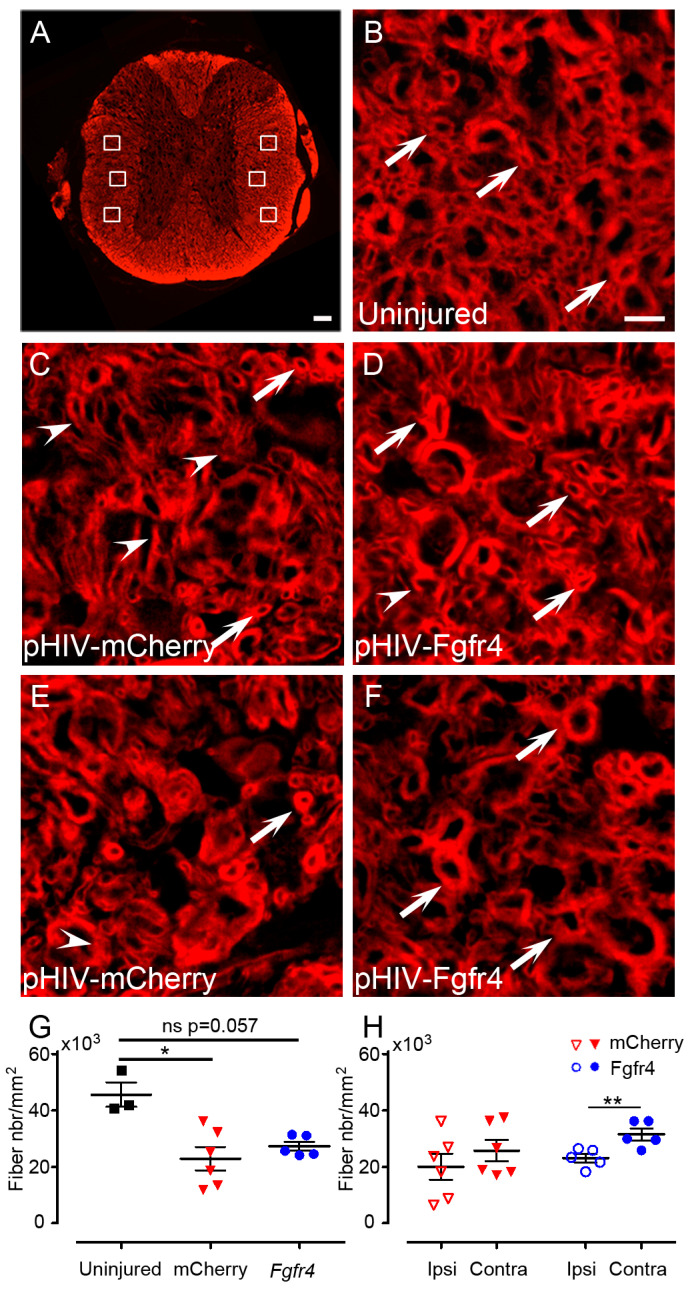
Quantification of spared myelin after SCI and pHIV-Fgfr4 injections. Axial section of a mouse spinal cord stained with fluoromyelin (A). White boxes indicate locations of the 6 high magnification images acquired per sections for quantifications of spared myelin fibers (A). Representative image of the white matter in an uninjured mouse (B). Representative images of the white matter in pHIV-mCherry (C,E), and pHIV-Fgfr4 (D,F) animals at 5 mm caudal to the lesion. Representative images on the ipsilateral (C,D) and contralateral (E,F) side of the lesion. Arrowheads point to damaged myelin fibers (uncomplete myelin sheath; (C)–(E)) and arrow point to spared myelin fibers (complete myelin sheath (B)–(F)). Quantifications were performed on 1 section located 3 mm rostral and 1 section located 3 mm caudal to the lesion epicenter. Density of the overall spared myelinated fibers (rostral and caudal; ipsilateral and contralateral; equivalent age and locations matches were taken for uninjured) (G). Density of spared myelinated fibers (rostral and caudal) ipsilateral and contralateral to the lesion site (H). Data from uninjured animals (black), animals that received the experimental vector (blue), and the control vector (red) are represented. Data are expressed as mean per mouse ± SEM. Student’s un-paired t-test: * p ≤ 0.05 (G) and paired t (H) ** p ≤ 0.01. Number of female C57BL6/6J mice: 3 uninjured, 6 control (pHIV-mCherry), and 5 experimental (pHIV-Fgfr4). In both groups (pHIV-mCherry and pHIV-Fgfr4) spinal cords were analyzed 6 weeks after SCI and vector injections. Scale bars: 100 µm (A) and 50 µm (B–F).

