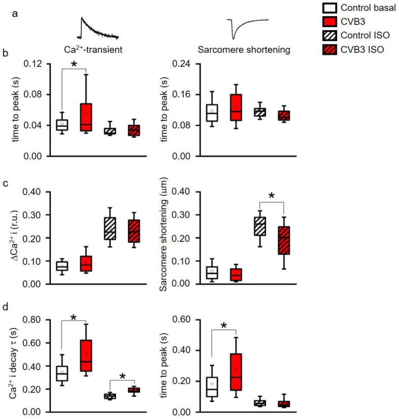
Figure 4.
(a) Time to peak of intracellular Ca2+ (Ca2+I) transients and of sarcomere shortening, (b) Ca2+-transient (ΔCa2+i) and sarcomere shortening amplitudes, (c) time constant τ of a single exponential decay function fitted to the decay phase of Ca2+-transients and to sarcomere relaxation phase. * indicates p < 0.05 vs. WT controls. All parameters recorded under acute stimulation with isoproterenol (ISO) were significantly altered vs. basal (p < 0.05, not indicated in the figure for clarity). (box: 25th–75th percentile, whiskers: 10th–90th percentile, horizontal line: median, square: mean). (d) decay of Ca2+ and time to peak were simultaneously measured and analyzed. Ca2+ decay time was significantly altered between control and CVB3-expressing cardiomyocytes under basal and stimulated conditions. Time to peak was only altered under basal conditions indicating a calcium-contraction uncoupling due to CVB3 expression (* indicates p < 0.05).

