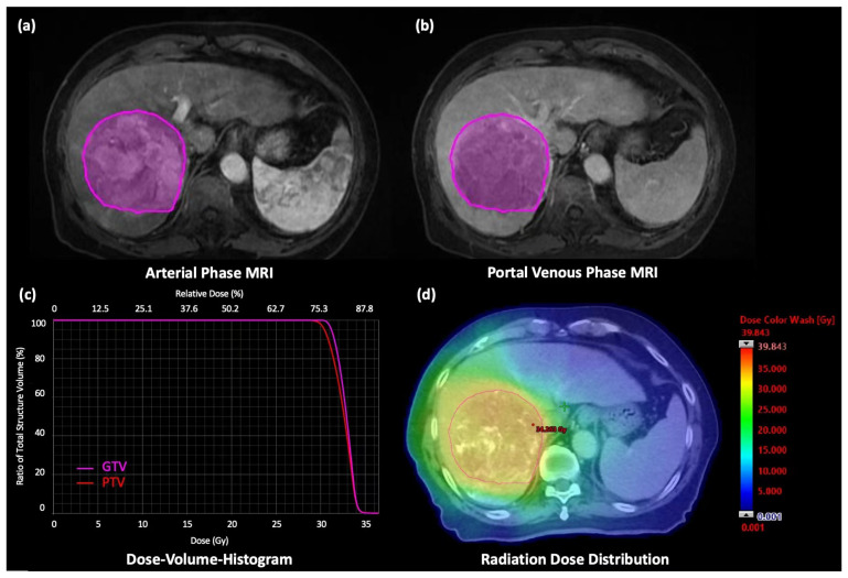
Figure 1.
A 76-year-old male patient was diagnosed with advanced-staged HCC. (a) Axial AP T1W MR image with the VOI, (b) Axial PVP T1W MR image with the VOI. SBRT of 30 Gy was prescribed in 5 fractions to the tumor. (c) DVH of gross tumor volume (GTV) and planning target volume (PTV) generated from the treatment planning system and (d) Dose distribution of the SBRT treatment plan.

