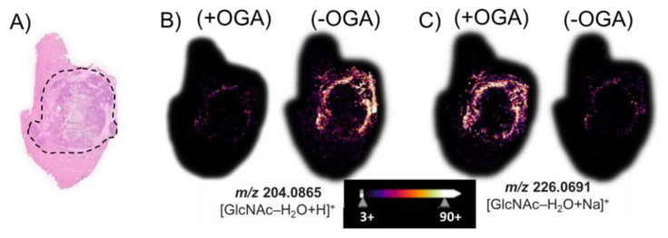
Figure 4.
(A) The H&E stain of a hepatic tumor section contains an outlined tumor region composed of necrotic tumor with viable tumor region found at the interface to the healthy tissue. MALDI imaging of (B) the water-loss oxonium ion of GlcNAc, m/z 204.0865; and (C) the sodium-cationized water-loss oxonium ion, m/z 226.0691. (+) indicates the use of OGA, while (−) indicates an untreated serial section. The tissue sections were treated with PNGase F to remove all N-glycans prior to the OGA (+/−) treatment.

