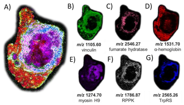
Figure 5.
MSI of peptide signatures observed after on-tissue PNGase F and OGA treatments and trypsin digestion (and matched to peptides characterized using in-situ gel trypsin LCMS/MS analysis that correspond to known proteins). (A) Co-registered collection of peptide matches. (B) m/z 1105.60 (−0.9 ppm error) corresponding to vinculin; (C) m/z 2546.27 (−6.5 ppm error) corresponding to fumarate hydratase; (D) m/z 1531.70 (−8.9 ppm error) corresponding to hemoglobin alpha; (E) m/z 1274.70 (−0.1 ppm error) corresponding to myosin heavy chain 9; (F) m/z 1786.87 (−6.1 ppm error) corresponding to ribose-phosphate diphosphokinase; and (G) m/z 2565.26 (6.2 ppm error) corresponding to aminoacyl tryptophan-tRNA synthase. The H&E stain of the same tissue section is shown in Figure S8 for reference and matched peptide sequences and protein identities are reported in Table S3.

