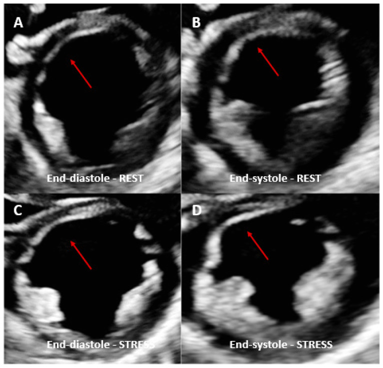
Figure 1.
Dobutamine stress echo performed in a patient with coronary artery vasculopathy (CAV). End-diastolic rest images (A) demonstrated a thinned anterior septal area compared to the other myocardial segments (red arrows) and not thickening during systole (B). Same view acquired at peak dobutamine stress (C,D) demonstrated no significant changes in the same area with no other regional wall motion abnormalities identified. Angiography was therefore performed, showing severe left anterior descending coronary artery stenosis.

