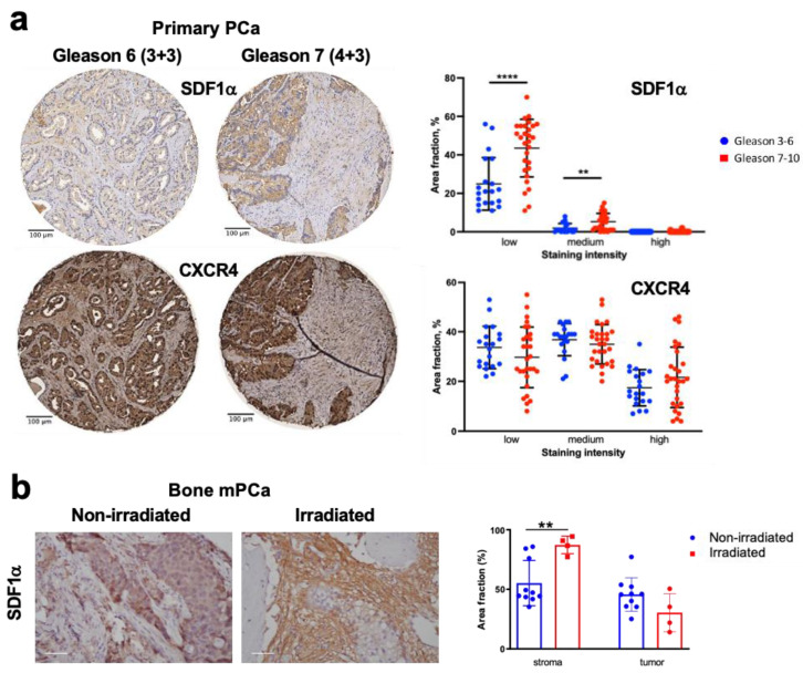
Figure 1.
Expression of SDF1α and CXCR4 in primary and bone metastatic prostate cancer (mPCa) tissues. (a) Representative images and quantitative analysis of SDF1α and CXCR4 immunostaining in primary PCa tissue, stratified by Gleason score (Gleason 3–6: n = 20, median score 6, median age 76 ± 9 years vs. Gleason 7–10: n = 29, median score 8, median age 78 ± 6 years) and staining intensity (high/medium/low) analyzed with ImageJ software using predefined thresholds for the respective categories. (b) Representative images and quantification of SDF1α expression by immunostaining in metastatic PCa tissue, radiation-naïve, or previously irradiated less than a month prior to surgery; scale bar: 50 µm. ** p < 0.01; **** p < 0.0001.

