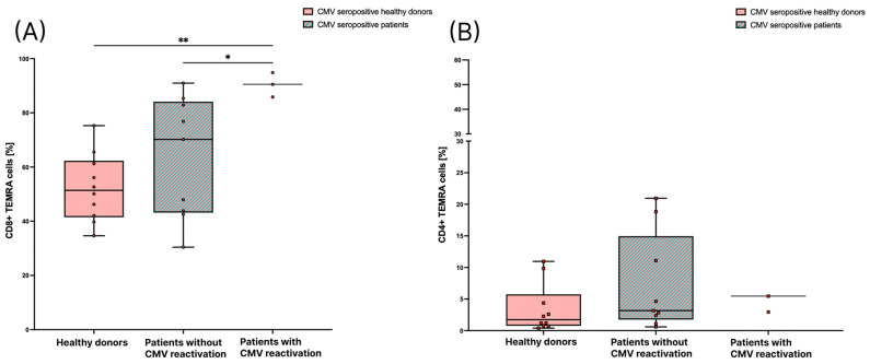
Figure 4.
Comparison of TEMRA cells in healthy donors and patients with or without CMV reactivation. TEMRA cells are depicted as percentages of CD8+ T cells in (A), and of CD4+ T cells in (B). TEMRA cells were measured in CMV seropositive healthy donors (n = 10), CMV seropositive patients before radiation therapy without reactivation (n = 9), and patients in the CMV seropositive group who had a reactivation of CMV (n = 3). For the statistical analysis, a non-parametric two-tailed Mann–Whitney U-test was applied (*: p < 0.05; **: p < 0.01).

