Figure 3.
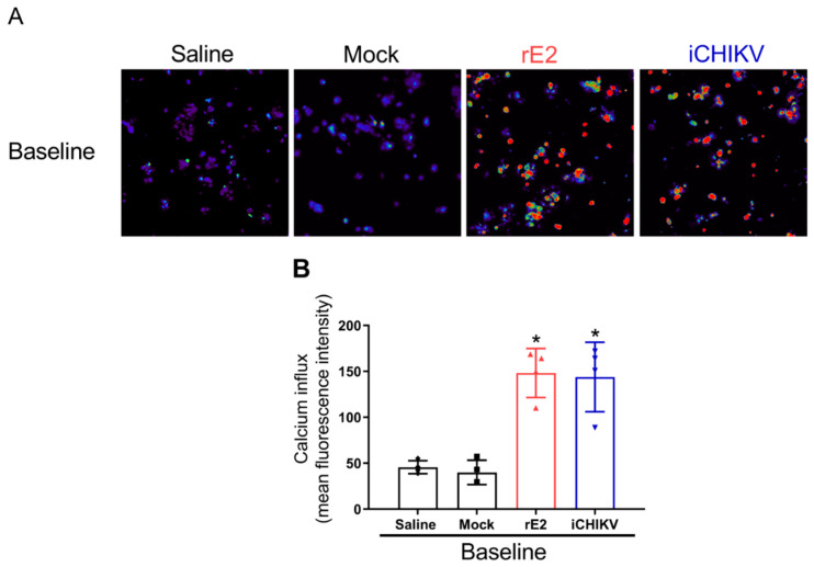
DRG neurons from iCHIKV and rE2-stimulated mice are activated. Seven hours after intra-articular (i.a.) injection of iCHIKV (100 FFU, 10 µL), rE2 (100 ng, 10 µL), saline or Mock (control, 10 µL), DRGs were dissected for calcium imaging using Fluo-4AM. Panel (A) displays representative fields of baseline fluorescence of DRG neurons dissected from saline or mock control mice and rE2 or iCHIKV-stimulated mice. Absent calcium levels are shown in blue, and increasing concentrations of calcium are shown by the change in color spectra up to red. Panel (B) shows the mean fluorescence intensity of calcium influx on the baseline (0-s mark). Image resolution: 288 × 220 mm (96 × 96 DPI). Results are expressed as mean ± SEM, n = 4 DRG plates (each plate is a neuronal culture pooled from six mice) per group per experiment, two independent experiments (* p < 0.05 vs. saline and mock; one-way ANOVA followed by Tukey’s post-test).

