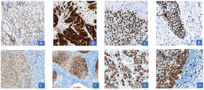
Figure 1.
Representative immunohistochemistry for tumoral PD-L1, P16, p53 and KI-67. Representative histology sections show a membranous tumoral PD-L1 expression (A), a positive nuclear and cytoplasmatic p 16 staining (B), a positive p53 tumor cell nuclei overexpression (C) and a strong KI-67 nuclear staining by immunohistochemistry (D); validated control sections according to ISO 17020: membranous PD-L1 expression (E), nuclear and cytoplasmatic staining of p 16 (F), p 53 nuclear overexpression (G), KI-67 nuclear and cytoplasmatic staining (H) by immunohistochemistry. Magnification 400×.

