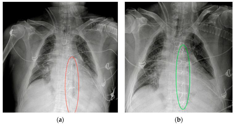
Figure 17.
Malpositioned IABP. (a) Anteroposterior CXR of a patient with an IAPB placed too distal from the aortic arch: the upper radiopaque mark can be seen at the level of the sixth intercostal space (red circle). (b) The same patient after repositioning of the IABP, with the upper mark now just under the aortic arch, in the proximal thoracic descending aorta, at the level of the fourth intercostal space (green circle).

