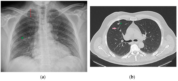
Figure 34.
Image of a CXR (a) obtained after CT-guided positioning (b) of a microcoil (green asterisk) into the lung parenchyma as a landmark to more easily recognize and resect the adjacent nodule (pink arrow) intraoperatively. Development of pneumothorax as a complication following this procedure is not uncommon [36], as it is possible to appreciate this in the reported CXR (red double arrow).

