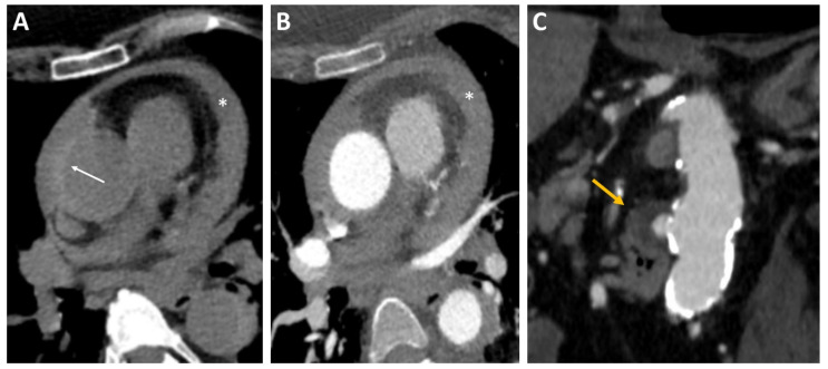
Figure 3.
Intramural hematoma on CT using (A) non-contrast CT and (B) CT angiography: crescentic, high-attenuating regions of eccentrically thickened aortic wall on non-contrast CT (arrow). A diffuse pericardial effusion (*) was also visible in both scans. (C) Penetrating ulcer on CT: CT angiography image showing a penetrating ulcer of the descending aorta as a contrast-filled, out-pouching into the thickened aortic wall (arrow). CT, computed tomography.

