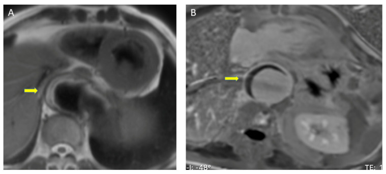
Figure 4.
Magnetic resonance imaging assessment of intramural hematoma (arrows). (A) Spin-Echo sequence. The eccentric thickening of the aortic wall has a high T1 signal, eliminating the possibility that it is an acute stage. (B) Late gadolinium- enhancement sequence. Aortic eccentric wall thickening with no mural enhancement, suggestive of intramural hematoma.

