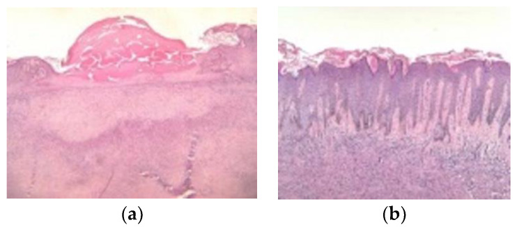
Figure 14.
(a) Histopathologic examination (hematoxylin and eosin, ×10): tumour proliferation localized in the papillary dermis and extending to the deep dermis, with interspersed collagen bundles, separated from the epidermis by a grenz zone. The overlying epidermis presents erosions centrally and collections in the keratin layer. (b) Histopathologic examination (hematoxylin and eosin, ×10): tumour proliferation composed of elongated and spindle-shape cells with elongated nuclei, in a fascicular-storiform configuration localized in the papillary dermis and extending to the deep dermis. The overlying epidermis has a hyperplastic appearance with hyperorthokeratosis, acanthosis, and elongation of the rate ridges. There is also follicular induction at the epidermis level.

