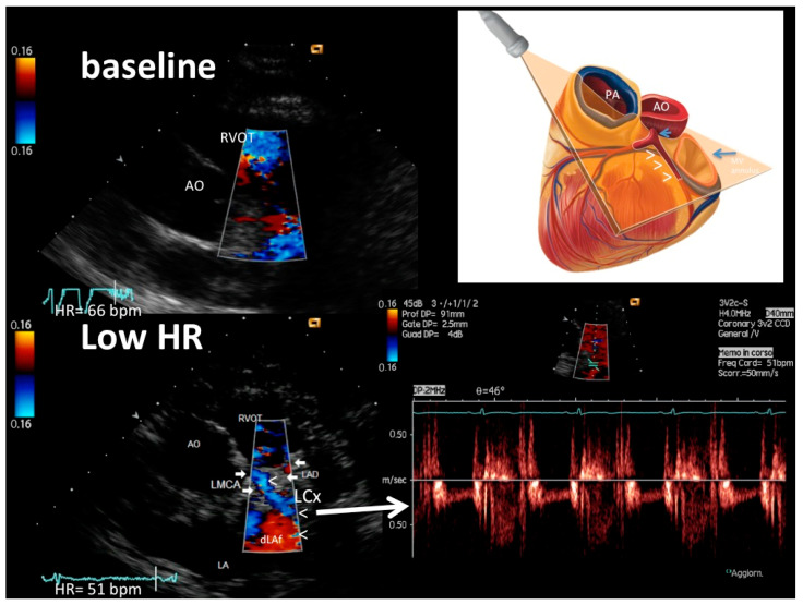
Figure 2.
An example of LMCA, proximal–mid LCx and proximal LAD color flow (plane orientation at the top right corner) at higher HR (66 b/m) (upper left) and after HR reduction (51 b/m) (bottom right): at the baseline with higher HR, no coronary flow is detected; after HR lowering (<60 b/m), optimal color signal is recorded in the LMCA (arrows), LCx (arrowheads) and very proximal LAD (arrows, signal in red); thanks to good color guidance optimal pulsed-wave Doppler quality in LCX is obtained (bottom right). The color signal is coded in blue both in LMCA and LCx and the LCx PW tracing is below the zero line because flow is away from the transducer. In the schematic cartoon (top right), the arrowheads indicate the LCx, the long arrow the mitral annulus and the short arrow the LMCA. LMCA = left main coronary artery color flow; LAD = proximal LAD color flow; LCx = proximal–mid left circumflex coronary artery color flow; RVOT = right ventricular outflow tract; pv = pulmonary valve; Ao = aorta; LA = left atrium.

