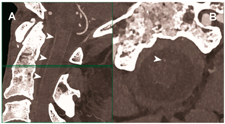
Figure 5.
Carotid CT angiography using photon-counting computed tomography. The figure shows advanced MIP reconstructions of a intracervical artery tree derived from a photo-counting CT (Scanner: NAEOTOM Alpha, Siemens) acquisition. In (A) a sagittal median view of the cervical region showing the course of the anterior spinal artery (normally not visible) in the ventral portion of the rachidial channel (arrowheads). In (B) the axial image at the level of the green plane showed on the left panel with the axial view of the anterior spinal artery (arrowhead).

