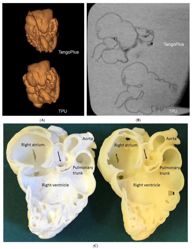
Figure 5.
Three-dimensional-printed CHD models with the use of different materials for comparison of model accuracy. (A): Three-dimensional CT volume rendering of the 3D-printed models showing similar anatomical details. (B): Two-dimensional axial CT views of the 3D printed models. (C): Inside view of cardiac chambers and aortic structures on both models. The white model is printed with TPU, while the yellow model is printed with TangoPlus. Arrows refer to the subaortic ventricular septal defect.

